Introduction
Cell adhesion is a complex process involved in embryogenesis, migration/invasion, tissue remodeling,
and wound healing. To perform these processes, cells adhere to extracellular matrix components (via
adhesion receptors), forming complexes with components of the cytoskeleton that ultimay affect cell
motility, differentiation, proliferation, and survival. The Cell Biolabs CytoSelect™ Cell Adhesion
Assay Kit provides a rapid, quantitative method for evaluating cell adhesion. The kit contains
sufficient reagents for the evaluation of 48 samples (40 Human Fibronectin-coated wells, 8 BSA-
coated wells).
Assay Principle
The CytoSelect™ Cell Adhesion Assay Kit utilizes a Fibronectin-coated 48-well plate (see Adhesion
Plate Layout below). First, cells are seeded onto the coated substrate, where the adherent cells are
captured. Next, unbound cells are washed away, and the adherent cells are fixed/stained. Finally, the
stain is extracted and quantified colorimetrically.
Related Products
1. CBA-051: CytoSelect™ 48-Well Cell Adhesion Assay (Fibronectin-Coated, Fluorometric Format)
2. CBA-052: CytoSelect™ 48-Well Cell Adhesion Assay (Collagen I-Coated, Colorimetric Format)
3. CBA-060: CytoSelect™ 48-Well Cell Adhesion Assay (Collagen IV-Coated, Colorimetric Format)
4. CBA-058: CytoSelect™ 48-Well Cell Adhesion Assay (Fibrinogen-Coated, Colorimetric Format)
5. CBA-056: CytoSelect™ 48-Well Cell Adhesion Assay (Laminin-Coated, Colorimetric Format)
6. CBA-070: CytoSelect™ 48-Well Cell Adhesion Assay (ECM Array, Colorimetric Format)
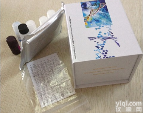
Kit Components
1. Fibronectin Adhesion Plate (Part No. 105001): One 48-well plate containing 40 Human
Fibronectin-coated wells and 8 BSA-coated wells (see layout below)
2. Cell Stain Solution (Part No. 11002): One Bottle – 10.0 mL
3. Extraction Solution (Part No. 11003): One Bottle – 10.0 mL
Adhesion Plate Layout
The layout below indicates the location of wells coated with Fibronectin and those coated with BSA.
2
A Fibronectin Fibronectin Fibronectin Fibronectin Fibronectin Fibronectin Fibronectin Fibronectin
B Fibronectin Fibronectin Fibronectin Fibronectin Fibronectin Fibronectin Fibronectin Fibronectin
C Fibronectin Fibronectin Fibronectin Fibronectin Fibronectin Fibronectin Fibronectin Fibronectin
D Fibronectin Fibronectin Fibronectin Fibronectin Fibronectin Fibronectin Fibronectin Fibronectin
E Fibronectin Fibronectin Fibronectin Fibronectin Fibronectin Fibronectin Fibronectin Fibronectin
F
BSA
BSA
BSA
BSA
BSA
BSA
BSA
BSA
Materials Not Supplied
1. Cell culture medium
2. Serum free medium, such as DMEM containing 0.5% BSA, 2 mM CaCl2 and 2 mM MgCl2
3. Cell culture incubator (37ºC, 5% CO2 atmosphere)
4. 1X PBS containing 2 mM CaCl2 and 2 mM MgCl2
5. Light microscope
6. 96-well microtiter plate
7. Microtiter plate reader
Storage
Store all kit components at 4ºC up to their expiration dates.
Assay Protocol
1. Under sterile conditions, allow the Fibronectin Adhesion Plate to warm up at room temperature
for 10 minutes.
2. Prepare a cell suspension containing 0.1-1.0 x 10⁶ cells/ml in serum free media. Agents that
inhibit or stimulate cell adhesion can be added directly to the cell suspension.
3. Add 150 µL of the cell suspension to the inside of each well (BSA-coated wells are provided as
a negative control).
4. Incubate for 30-90 min in a cell culture incubator.
3
5. Carefully discard or aspirate the media from each well (Note: Do not allow wells to dry).
Gently wash each well 4-5 times with 250 µL PBS.
6. Aspirate the PBS from each well and add 200 µL of Cell Stain Solution. Incubate for 10
minutes at room temperature.
7. Discard or aspirate the Cell Stain Solution from the wells. Gently wash each well 4-5 times
with 500 µL deionized water.
8. Discard the final wash and let the wells air dry.
9. Add 200 µL of Extraction Solution per well, and then incubate 10 minutes on an orbital shaker.
10. Transfer 150 µL from each extracted sample to a 96-well microtiter plate and measure the OD
560nm in a plate reader.
Example of Results
The following figures demonstrate typical results with the CytoSelect™ 48-Well Cell Adhesion Assay
Kit. One should use the data below for reference only. This data should not be used to interpret actual
results.
2.5
MDA-231
HT-1080
HEK293
2
1.5
1
0.5
0
BSA Col I Col IV FN LN VN
Figure 1. ECM-mediated Cell Adhesion. Serum starved cells were allowed to attach to ECM-
coated 48-well plate for 1 hr at 100,000 cells/well. Adherent cells were stained (left panel picture) and
quantified at OD 560nm after extraction (right panel figure).
References
1. Hynes, R. O. (1992) Cell 69:11-25.
2. Schwartz, M. A., Schaller, M. D. and Ginsberg, M. H. (1995) Annu. Rev. Cell Dev. Biol. 11:549-
599.
4
Recent Product Citations
1. Cervera, A.M. et al. (2008). Cells silenced for SDHB expression display characteristic features of
the tumor phenotype. Cancer Res. 68:4058-4067.
Warranty
These products are warranted to perform as described in their labeling and in Cell Biolabs literature when used in
accordance with their instructions. THERE ARE NO WARRANTIES THAT EXTEND BEYOND THIS EXPRESSED
WARRANTY AND CELL BIOLABS DISCLAIMS ANY IMPLIED WARRANTY OF MERCHANTABILITY OR
WARRANTY OF FITNESS FOR PARTICULAR PURPOSE. CELL BIOLABS’ sole obligation and purchaser’s
exclusive remedy for breach of this warranty shall be, at the option of CELL BIOLABS, to repair or replace the products.
In no event shall CELL BIOLABS be liable for any proximate, incidental or consequential damages in connection with the
products.
2004-2008: Cell Biolabs, Inc. - All rights reserved. No part of these works may be reproduced in any form without
permissions in writing.


 大鼠树突状细胞表面特异性C型凝集素-细胞间黏附分子3结合非整合素分子(DC-SIGN/CD209)酶联免疫吸附测定试剂盒Rat DC-SIGN/CD209 (DC Specific Intercellular Adhesion Molecule 3-Grabbing Nonintegrin) ELISA Kit
 MAdCAM-1(Human mucosal addressin cell adhesion molecule-1) ELISA Kit 人黏膜地址素细胞黏附分子
 ELISA. , 人黏膜地址素细胞黏附分子(MAdCAM-1)ELISA Kit, Human mucosal addressin cell adhesion molecule-1,MAdCAM-1 ELISA Kit
 Human mucosal addressin cell adhesion molecule-1,MAdCAM-1 ELISA Kit人黏膜地址素细胞黏附分子(MAdCAM-1)ELISA Kit
 人黏膜地址素细胞黏附分子(MAdCAM1)elisa试剂盒Human MAdCAM1 (Mucosal Addressin Cell Adhesion Molecule 1) ELISA Kit
 Human mucosal addressin cell adhesion molecule-1,MAdCAM-1 ELISA Kit人黏膜地址素细胞黏附分子(MAdCAM-1)ELISA试剂盒
 白细胞激活黏附因子(ALCAM)检测试剂盒(酶联免疫吸附试验法)CD166; MEMD; Activated Leucocyte Cell Adhesion Molecule
 人白细胞活化黏附因子(ALCAM)ELISA试剂盒哪个厂家好,人白细胞活化黏附因子(ALCAM)ELISA试剂盒优惠价,人白细胞活化黏附因子(ALCAM)ELISA试剂盒现货
 Rat DC specific intercellular adhesion molecule 3-grabbing nonintegrin,DC-SIGN/CD209 ELISA Kit大鼠树突状细胞表面特异性C型凝集素-细胞间黏附分子3结合非整合素分子(DC-SIGN/CD209)
 CBA-211 CytoSelect™ 96-well Leukocyte-epithelium Adhesion Kit CytoSelect™96孔白细胞,上皮细胞黏附试剂盒


