
品牌 其他品牌 供货周期 现货
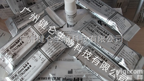
创仑乙型流感病毒IgM核酸快速检测试纸条
广州健仑生物科技有限公司
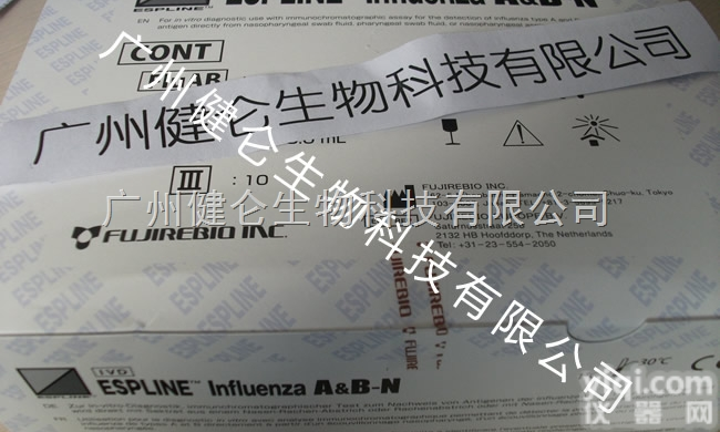
广州健仑长期供应各种流感检测试剂,包括进口和国产的品Pai,主要包括日本富士瑞必欧、日本生研、美国BD、美国NovaBios、美国binaxNOW、凯必利、广州创仑等主流品Pai。
创仑乙型流感病毒IgM核酸快速检测试纸条
我司还提供其它进口或国产试剂盒:登革热、疟疾、流感、A链球菌、合胞病毒、腮病毒、乙脑、寨卡、黄热病、基孔肯雅热、克锥虫病、违禁品滥用、肺炎球菌、军团菌、化妆品检测、食品安全检测等试剂盒以及日本生研细菌分型诊断血清、德国SiFin诊断血清、丹麦SSI诊断血清等产品。
欢迎咨询
欢迎咨询2042552662


想了解更多的产品及服务请扫描下方二维码:

【公司名称】 广州健仑生物科技有限公司
【市场部】 杨永汉
【】
【腾讯 】 2042552662
【公司地址】 广州清华科技园创新基地番禺石楼镇创启路63号二期2幢101-103室

大多数通过仔细询问病史可以粗略明确诊断方向,根据辅助检查可明确诊断。所以重视病史询问是一个非常重要的内容。比如询问:什么时候发病、发病的缓急、持续时间,发绀的部位,严重程度,是否对称性发绀。有无其他伴随症状,比如呼吸困难、咳嗽、咳痰、咯血、心慌、气促、胸闷、胸痛、恶心、呕吐等。有无水肿,水肿的部位,是否对称性等。有无接触某些化学药物病史。既往身体状况如何,有无心、肺疾病史等。
体格检查
体格检查一般跟收集病史同时进行。结合相应病史,观察发绀的各个信息点,比如紫绀部位、局部皮肤温度、有无杵状指、心肺疾病体征等。
辅助间一般是明确诊断所必须的。可以根据病史及体格检查所收集的信息,加以筛选地进行某些必要的辅助检查。比如胸片、心电图、超声心动图等,若怀疑有心脏方面疾病,可以选择更为复杂的辅助检查,比如心导管检查、选择性血管造影等。怀疑肺部疾病,则血气分析、肺功能检查等应该进行。怀疑下肢静脉狭窄或栓塞,则肢体血管的多普勒检查就很必要。其发生机制是由于大血管间存在异常通道,部分静脉血未通过肺进行氧合作用即经异常通道混入体循环动脉血中,如分流量超过心排血量的1/3即可引起紫绀。常见的疾病包括法洛四联症、大血管转位、永存动脉干、完全性肺静脉异位引流等。
非紫绀型先天性心脏病
非紫绀型先天性心脏病后期如出现继发性肺动脉高压即艾森门格综合征(Eisenmenger综合征),出现右向左分流,亦可出现紫绀,如室间隔缺损、动脉导管未闭、房间隔缺损、肺动脉狭窄及左心发育不良综合征等。
心胸手术后并发症
如引起右向左分流,亦可引起紫绀。对于心血管手术后的患者,如出现ZX性紫绀,在除外肺部疾患及心脏功能不全的情况下,应想到心血管手术部位或其它部位右向左分流的可能,其特点是二维UCG或心血管造影可见异常的血流通道。
其 他
如各种原因引起持续的快速型心律失常,可引起周围皮肤青紫,其中包括起搏器介导的快速心律失常,心电图可有快速心律失常的表现。另外,心脏原发肿瘤,有时由于血液回流不畅,亦可引起紫绀,如心房黏液瘤和右心室纤维瘤,其主要症状是体循环栓塞、发热、关节痛等,由于其病理生理特点主要是间歇性的肺血流量改变或周围静脉回流障碍,与体位有关,因此听诊常有变化,行心血管造影可见心腔内充盈缺损,而超声心脏检查可见心腔内肿块。
Mostly by carefully asking the medical history can be a rough diagnosis of the direction, according to the auxiliary examination can confirm the diagnosis. So pay attention to medical history is a very important content. For example, ask: when the onset, onset of urgency, duration, location of cyanosis, severity, whether the symmetry of cyanosis. Is there any other accompanying symptoms, such as dyspnea, cough, expectoration, hemoptysis, palpitation, shortness of breath, chest tightness, chest pain, nausea, vomiting. Whether or not edema, edema, symmetry and so on. Have any contact with certain medical history. Past physical condition, inadvertent, lung disease history.
Physical examination
Physical examination with the general collection of medical history at the same time. Combined with the corresponding history, to observe the cyanosis of various information points, such as cyanosis site, the local skin temperature, with or without clubbing, cardiopulmonary disease signs.
Auxiliary room is usually a clear diagnosis of the necessary. According to medical history and physical examination collected information, to be screened for some necessary auxiliary examination. Such as chest X-ray, ECG, echocardiography, etc., if there is suspicion of heart disease, you can choose more complicated auxiliary examinations, such as cardiac catheterization, selective angiography. Suspected lung disease, the blood gas analysis, pulmonary function tests should be carried out. Doubt of venous stenosis or embolism, the limb vascular Doppler examination is necessary. The mechanism is due to the presence of abnormal channels between major blood vessels, part of the venous blood without oxygenation through the lung that is mixed with systemic arterial blood through abnormal channels, such as subluxation volume of cardiac output of more than 1/3 can cause cyanosis. Common diseases include tetralogy of Fallot, major vessel transposition, permanent artery dry, complete pulmonary venous drainage and so on.
Non-cyanotic congenital heart disease
Non-cyanotic congenital congenital heart disease secondary to secondary pulmonary hypertension or Eisenmenger syndrome (Eisenmenger syndrome), the right to left shunt may also be cyanotic, such as ventricular septal defect, patent ductus arteriosus, atrial Septal defect, pulmonary stenosis and left heart hypoplasia syndrome.
Complications after thoracotomy
Such as causing right to left shunt, can also cause cyanosis. For patients after cardiovascular surgery, such as the emergence of central cyanosis, in addition to lung disease and cardiac insufficiency, should think of cardiovascular surgery or other parts of the right to left shunt may be characterized by two-dimensional UCG or Cardiovascular angiography shows abnormal blood flow channels.
Other
Prolonged tachyarrhythmias, such as various causes, can cause bruising on the surrounding skin, including pacemaker-mediated tachyarrhythmias and electrocardiogram with tachyarrhythmia. In addition, primary cardiac tumors, sometimes due to poor blood reflux, can also cause cyanosis, such as atrial myxoma and right ventricular fibroma, the main symptoms are systemic embolism, fever, joint pain, etc., due to its pathophysiological characteristics are intermittent Sexual changes in pulmonary blood flow or peripheral venous reflux disorders, and body position, so often changes in auscultation, cardiovascular cardioveal filling filling defects can be seen, and intracardiac echocardiography can be seen intraluminal mass.
 荧光PCR 创仑乙型流感病毒IgM核酸快速检测试纸条
 美国binaxNOW 乙型流感病毒Victoria核酸荧光探针检测试纸
 乙型流感检测卡 乙型流感试纸 乙型流感半成品 乙型流感胶体金大板
乙型流感试纸检测 B型流感ELISA检测试纸
乙型流感检测卡 乙型流感试纸 乙型流感半成品 乙型流感胶体金大板
乙型流感试纸检测 B型流感ELISA检测试纸
 诺瓦克试纸 诺如病毒核酸检测试剂盒(核酸pcr)
 德国进口SENOVA 埃博拉病毒核酸检测试纸(核酸PCR)
 流感试纸 甲乙型流感病毒试纸
流感AB病毒试纸 流感甲乙型病毒联检试纸
购买流感试纸 甲乙型流感检测试纸
流感试纸 甲乙型流感病毒试纸
流感AB病毒试纸 流感甲乙型病毒联检试纸
购买流感试纸 甲乙型流感检测试纸
 结核分枝杆菌核酸检测试剂盒(恒温扩增-试纸条法)
 检测试纸 支气管炎甲型乙型流感病毒诊断试剂盒
48T/96T 人乙型肝炎病毒脱氧核糖核酸ELISA试剂盒说明书
检测试纸 支气管炎甲型乙型流感病毒诊断试剂盒
48T/96T 人乙型肝炎病毒脱氧核糖核酸ELISA试剂盒说明书
本产品信息由(广州健仑生物科技有限公司)为您提供,内容包括(荧光PCR 创仑乙型流感病毒IgM核酸快速检测试纸条)的品牌、型号、技术参数、详细介绍等;如果您想了解更多关于(荧光PCR 创仑乙型流感病毒IgM核酸快速检测试纸条)的信息,请直接联系供应商,给供应商留言。若当前页面内容侵犯到您的权益,请及时告知我们,我们将马上修改或删除。


