
品牌 其他品牌 供货周期 现货
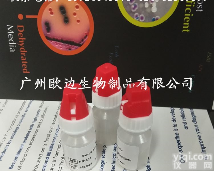
ACTH促肾上腺皮质激素(兔多克隆抗体)
广州健仑生物科技有限公司
ACTH 即促肾上腺皮质激素(adreno-cortico-tropic-hormone)是维持肾上腺正常形态和功能的重要激素。它的合成和分泌是垂体前叶在下丘脑促皮质素释放激素(CRH)的作用下,在腺垂体嗜碱细胞内进行的。此抗体与人的ACTH反应,同时与多种哺乳动物的ACTH有交叉反应,可用于垂体腺瘤的功能性分类,有助于区分原发性和转移性垂体肿瘤。嗜铬细胞瘤等部分神经内分泌肿瘤也可出现阳性反应。
我司还提供其它进口或国产试剂盒:登革热、疟疾、流感、A链球菌、合胞病毒、腮病毒、乙脑、寨卡、黄热病、基孔肯雅热、克锥虫病、违禁品滥用、肺炎球菌、军团菌、化妆品检测、食品安全检测等试剂盒以及日本生研细菌分型诊断血清、德国SiFin诊断血清、丹麦SSI诊断血清等产品。
欢迎咨询
欢迎咨询
【产品介绍】
细胞定位:细胞浆
适用组织:石蜡/冰冻
阳性对照:垂体
抗原修复:热修复(柠檬酸)
抗体孵育时间:30-60min
| 产品编号 | 产品名称 | 克隆型别 |
| OB001 | AACT(抗胰糜蛋白酶) | polyclonal |
| OB002 | AAT(抗胰蛋白酶) | polyclonal |
| OB003 | ACTH(促肾上腺皮质激素) | polyclonal |
| OB004 | Actin,Muscle Specific(肌肉特异性肌动蛋白) | HHF35 |
| OB005 | Actin,Smooth Muscle(平滑肌肌动蛋白) | 1A4 |
| OB006 | AFP(甲胎蛋白) | polyclonal |
ACTH促肾上腺皮质激素(兔多克隆抗体)
1、传染源:慢性病人、恢复期病人及健康的“排包囊者”为本病的传染源。急性病人,当其粪便中仅排出滋养体时,不是传染源。
2、传播途径:包囊在土壤中可以生存8天以上;在潮湿及凉爽环境内,如粪便中可以生存几个星期。包囊可以通过污染饮水、食物、蔬菜等进入人体。在卫生环境恶劣的地方,水源或食物易被粪便所污染。在以粪便作肥料的地区,未洗净、未煮熟的蔬菜是重要的传播因素。 蝇类及蟑螂都可接触粪便,体表携带和呕吐排便,将包囊污染食物而成为重要传播媒介。
3、流行特征:溶组织内阿米巴病在热带、亚热带、温带地区,发病较多,以秋季为多,夏季次之。发病率农村高于城市,男子多于女子,成年多于儿童,幼儿患者很少,可能与吞食含包囊食物机会的多少有关。
病理改变
结肠病变以局限性粘膜下小脓肿开始,其孤立散在分布。组织破坏逐步向纵深发展,自粘膜下层直至肌层,形成口小底大的典型烧瓶样溃疡。早期病变仅可见粘膜小溃疡,表面周围略上翻,但边缘不整齐,溃疡表面可见深黄色或灰黑色坏死组织,在其深部可找到滋养体。溃疡与溃疡之间的粘膜一般正常,如无继发细菌感染,则无炎症反应。病变主要属于组织坏死、细胞溶化的性质,而非炎症。
溃疡底部的血管有血栓形成,但有时病变可破坏小动脉酿成严重甚至危及生命的出血。溃疡亦可穿破肌层直至肠壁,使后者变得极薄,肠内容可以渗漏至腹腔,或穿破肠壁,造成局限性腹腔脓肿或弥漫性腹膜炎。
在慢性病变中,息肉样残片可伸向肠腔。溃疡愈合后仍可见到疤痕痕迹。由于滋养体反复侵入粘膜,加以细菌继发感染,肠粘膜组织增生肥大,可出现大块状肉芽肿,成为阿米巴瘤(ameboma),其多见于抗原抗体、抗原抗体直肠交接处、横结肠及盲肠。阿米巴瘤有时极为巨大、质硬,难以同大肠癌肿鉴别。
阿米巴病变部位的分布依次为盲肠与升结肠、抗原抗体、直肠、阑尾和回肠下段。滋养体可进入门静脉血流,在肝内形成脓肿,且可以栓子形式流入肺、脑、脾等组织与抗原抗体,形成脓肿。
病变在显微镜下所见,组织坏死系其主要变化。轻度或中度淋巴细胞浸润,并伴有少量中性粒细胞,此在有细菌继发感染时更著。阿米巴滋养体满布于整个病损中,尤其多见于病损扩展的边缘,甚至在邻近的正常组织中也有。
我司还提供其它进口或国产试剂盒:登革热、疟疾、流感、A链球菌、合胞病毒、腮病毒、乙脑、寨卡、黄热病、基孔肯雅热、克锥虫病、违禁品滥用、肺炎球菌、军团菌、化妆品检测、食品安全检测等试剂盒以及日本生研细菌分型诊断血清、德国SiFin诊断血清、丹麦SSI诊断血清等产品。
想了解更多的产品及服务请扫描下方二维码:

【公司名称】 广州健仑生物科技有限公司
【市场部】 杨永汉
【】
【腾讯 】
【公司地址】 广州清华科技园创新基地番禺石楼镇创启路63号二期2幢101-103室


1, the source of infection: chronic patients, convalescent patients and healthy "platoon" as the source of infection. Acute patients are not the source of infection when only their trophozoites are excreted.
2, route of transmission: cysts in the soil can survive for more than 8 days; in humid and cool environment, such as the stool can survive for a few weeks. Encapsulation can enter the body by contaminating drinking water, food, vegetables and the like. In poor sanitary conditions, water or food is easily contaminated by faeces. In areas where manure is used as fertilizer, unwashed, uncooked vegetables are important sources of transmission. Flies and cockroaches can be exposed to excrement, body surface and vomiting, defecation, the capsule contaminated food has become an important media.
3, epidemiological characteristics: Entamoeba histolytica in tropical, subtropical, temperate regions, more incidence, with more fall, summer followed. Morbidity is higher in rural areas than in urban areas, with more males than females, more adulthood than children, and very few toddlers, which may be related to the chance of swallowed cysts containing food.
Pathological changes
Colon lesions start with localized submucosal small abscesses that are scattered in isolation. Tissue destruction gradually developed in depth, from the submucosa until the muscularis, the formation of large typical small mouth ulcers flask. Early lesions only visible mucosal ulceration, the surface slightly upside down, but the edge is not neat, the surface of the ulcer can be seen dark yellow or gray-black necrosis in the deep can find trophozoites. The mucosa between the ulcer and the ulcer is generally normal, without secondary bacterial infection, there is no inflammatory reaction. Lesions mainly belong to the nature of tissue necrosis, cytolysis, rather than inflammation.
The bottom of the ulcer blood vessels thrombosis, but sometimes the lesion can damage the small arteries causing serious and even life-threatening bleeding. Ulcers can also break through the muscular layer until the intestinal wall, making the latter extremely thin, intestinal contents can leak to the abdominal cavity, or wear broken bowel wall, resulting in localized abdominal abscess or diffuse peritonitis.
In chronic lesions, polypoid fragments can stretch to the intestine. Can still be seen after ulcer healing scar marks. As the trophozoites repeatedly invade the mucosa, bacterial secondary infection, intestinal mucosal hyperplasia hypertrophy, there may be massive granulomas, become ameboma (ameboma), which more common in antigen and antibody, antigen and antibody rectal junction, transverse colon and Cecum. Amoeba tumor is sometimes extremely large, hard, difficult to identify with colorectal cancer.
The distribution of the amoebic lesions was caecal and ascending colon, antigen antibody, rectum, appendix and lower ileum. Trophoblast can enter the portal vein blood flow, forming an abscess in the liver, and can flow into the lungs, brain, spleen and other tissue and antigen antibodies in the form of emboli to form abscesses.
Lesions seen under the microscope, tissue necrosis of its main changes. Mild or moderate lymphocytic infiltration, with a small amount of neutrophils, is more pronounced with bacterial secondary infection. Amoeba trophozoites are found throughout the entire lesion, most often at the margin of lesion expansion, even in adjacent normal tissues.
 ACTH促肾上腺皮质激素(兔多克隆抗体)
 去甲肾上腺素转运蛋白/神经递质去甲肾上腺素转运体抗体,Anti-NET1抗体
 Anti-NET1抗体,去甲肾上腺素转运蛋白/神经递质去甲肾上腺素转运体抗体价格
 Anti-NET1抗体,去甲肾上腺素转运蛋白/神经递质去甲肾上腺素转运体抗体价格
 Biotin 标记NET1抗体,去甲肾上腺素转运蛋白/神经递质去甲肾上腺素转运体抗体
 NET1抗体,去甲肾上腺素转运蛋白/神经递质去甲肾上腺素转运体抗体FITC.PE
 α1肾上腺素能受体抗体低价促销,α1肾上腺素能受体抗体价格
 alpha-2肾上腺素能受体抗体批发,alpha-2肾上腺素能受体抗体直销
alpha-2肾上腺素能受体抗体规格,北京alpha-2肾上腺素能受体抗体
肾上腺素能受体抗体规格,北京肾上腺素能受体抗体
alpha-2肾上腺素能受体抗体批发,alpha-2肾上腺素能受体抗体直销
alpha-2肾上腺素能受体抗体规格,北京alpha-2肾上腺素能受体抗体
肾上腺素能受体抗体规格,北京肾上腺素能受体抗体
 AP,APC,FITC NET1抗体,去甲肾上腺素转运蛋白/神经递质去甲肾上腺素转运体抗体Go...
 NET1抗体 去甲肾上腺素转运蛋白/神经递质去甲肾上腺素转运体抗体
本产品信息由(广州欧边生物制品有限公司)为您提供,内容包括(ACTH促肾上腺皮质激素(兔多克隆抗体))的品牌、型号、技术参数、详细介绍等;如果您想了解更多关于(ACTH促肾上腺皮质激素(兔多克隆抗体))的信息,请直接联系供应商,给供应商留言。若当前页面内容侵犯到您的权益,请及时告知我们,我们将马上修改或删除。

沪公网安备 31011502008050号


