品牌 其他品牌 供货周期 现货
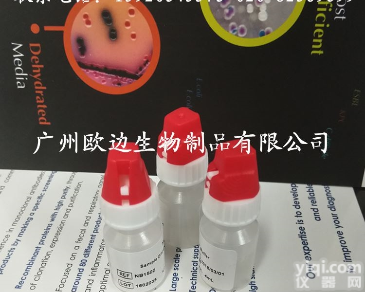
芬兰流感胶体金检测卡
广州健仑生物科技有限公司
广州健仑长期供应各种PCR试剂盒,主要代理进口和国产品Pai的流行病毒PCR检测试剂盒。例如:甲乙型流感病毒核酸检测试剂盒、黄热病毒核酸检测试剂盒、诺如病毒核酸检测试剂盒、登革病毒核酸检测试剂盒、基孔肯雅病毒核酸检测试剂盒、结核杆菌核酸病毒检测试剂盒、孢疹病毒核算检测试剂盒、西尼罗河病毒PCR检测试剂盒、呼吸道合胞病毒核酸检测试剂盒、冠状病毒PCR检测试剂盒等等。虫媒体染病系列、呼吸道病原体系列、发热伴出疹系列、消化道及食源感染系列。
广州健仑长期供应各种流感检测试剂,包括进口和国产的品Pai,主要包括日本富士瑞必欧、日本生研、美国BD、美国NovaBios、美国binaxNOW、英国clearview、凯必利、广州创仑等主流品Pai。
主要检测:甲型流感病毒检测试剂、乙型流感病毒检测试剂、甲乙型流感病毒检测试剂、A+B流感病毒检测试剂盒、流感病毒抗原快速检测卡、流感病毒抗体快速检测试剂盒、流感快速检测试剂 c1c2。
芬兰流感胶体金检测卡
1、推荐采集的呼吸道标本种类
疾病发病后应尽快采集如下标本:鼻拭子、咽拭子、鼻腔吸取物、鼻腔冲洗液。气管插管的病人也应收集气管吸取物。标本应置于无菌病毒采样液,并立即用冰块或冰排保存或置于4 ℃ (冰箱),并马上送至实验室。(临床样品采集人员及实验室检测人员的感染控制指导请见相关文件。) 采样同时填写疑似人感染甲型H1N1感染病例标本采样单。
2、拭子的选择
标本应使用头部为合成纤维的拭子(例如,聚酯纤维),用铝或塑料做柄。不推荐棉拭子和木柄。标本采集管应包含3毫升病毒采样液(含有蛋白质稳定剂,阻止细菌和真菌生长的抗生素,缓冲液)
3、临床标本储存要求
保存在4℃(不能超过4天)或者-70℃或-70℃以下。有条件的实验室均应保存在-70℃或-70℃以下。不应保存在-20℃。
4、标本的分装处理
标本送至实验室后,立即进行处理,避免反复冻融。将原始标本分为三份,一份用于核酸检测,一份用于病毒分离,一份保存待复核。
5、标本的运输
疑似人感染甲型H1N1感染病例列为 A类,用UN2814包装运输。填写疑似人感染甲型H1N1感染病例标本送检单。
这种

我司还提供其它进口或国产试剂盒:登革热、疟疾、流感、A链球菌、合胞病毒、腮病毒、乙脑、寨卡、黄热病、基孔肯雅热、克锥虫病、违禁品滥用、肺炎球菌、军团菌、化妆品检测、食品安全检测等试剂盒以及日本生研细菌分型诊断血清、德国SiFin诊断血清、丹麦SSI诊断血清等产品。
欢迎咨询
欢迎咨询
想了解更多的产品及服务请扫描下方二维码:

【公司名称】 广州健仑生物科技有限公司
【市场部】 欧
【】
【腾讯 】
【公司地址】 广州清华科技园创新基地番禺石楼镇创启路63号二期2幢101-103室


4. J. D. Robertson 1959 用超薄切片技术获得了清晰的细胞膜照片,显示暗-明-暗三层结构,厚约
7.5nm。这就是所谓的“单位膜”模型。它由厚约3.5nm的双层脂分子和内外表面各厚约2nm的蛋白质构成
。单位膜模型的不足之处在于把膜的动态结构描写成静止的不变的。
5. S. J. Singer & G. Nicolson 1972 根据免疫荧光技术、冰冻蚀刻技术的研究结果,在”单位膜”
模型的基础上提出”流动镶嵌模型”。强调膜的流动性和膜蛋白分布的不对称性,[2] 但并不能说明具
有流动性的质膜在变化过程中怎样保持膜的相对完整性和稳定性。
6. Wallach 1975 提出“晶格镶嵌模型”。认为,膜蛋白对脂类分子的运动有限制作用,镶嵌蛋白和其
周围的脂类分子形成膜中晶态部分,而具有流动性的脂类呈小片的点状分布。因此,脂类的流动性是局
部的,并非整个脂类双分子都在进行流动,这就比较合理地解释了生物膜既具有流动性又可以保持相对
完整性和稳定性的原因[3] 。
7.Jain &White 1977 提出“板块镶嵌模型”。认为在流动的脂双分子层中存在许多大小不同、刚性较大
的能独立移动的脂类板块,这些有序结构板块间存在流动的脂类 ,二者处于连贯的动态平衡[3] 。
6. 1988年,提出“脂筏模型”。即脂双分子层并不是一个完全均匀的二维流体,内部存在富含胆固醇、
鞘脂和特定种类膜蛋白的微区,这一区域较厚且较少流动,被称为“脂筏”;其如同一个蛋白质停泊的
平台,与膜的信号转导、蛋白质分选均有密切的关系[4] 。
化学组成编辑质膜主要由膜脂和膜蛋白组成,另外还有少量糖,主要以糖脂和糖蛋白的形式存在。膜脂是膜的基本骨
架,膜蛋白是膜功能的主要体现者。动物细胞膜通常含有等量的脂类和蛋白质。
质膜的流动性对生物体有很大影响。温度、ph、脂分子结构、胆固醇等多方面影响流动性。古生菌的质
膜是由单层或单双层交替的磷脂分子组成,因此流动性较差,抗逆性强。是构成膜脂的基本成分,约占
整个膜脂的50%以上。磷脂分子的主要特征:
具有一个极性头和两个非极性的尾(脂肪酸链),但存在于线粒体内膜和某些细菌质膜上的心磷脂具有4。
4. J. D. Robertson 1959 A clear picture of the cell membrane was obtained using the ultrathin sectioning technique showing a dark-bright-dark three-layer structure with a thickness of about
7.5 nm. This is the so-called "unit membrane" model. It consists of bilayer lipid molecules about 3.5 nm in thickness and proteins each about 2 nm thick on the inner and outer surfaces
. The disadvantage of the unit membrane model is that the dynamic structure of the membrane is described as static and constant.
5. S. J. Singer & G. Nicolson 1972 According to the results of immunofluorescence and freeze-etching techniques,
Based on the model proposed " mosaic model." Emphasize membrane fluidity and membrane protein distribution asymmetry, [2] but it does not explain
The fluidity of the plasma membrane in the process of how to maintain the relative integrity and stability of the membrane.
6. Wallach 1975 proposed "lattice mosaic model." It is thought that the membrane proteins have a limiting effect on the movement of lipid molecules, the mosaic proteins and their
The surrounding lipid molecules form the crystalline portion of the membrane, while the flowable lipids are distributed in small patches. Therefore, the mobility of lipids is a bureau
Department, not the entire lipid bimolecular are in the flow, which is more reasonable to explain the biofilm has both fluid and can maintain relative
Integrity and stability of the reasons [3].
7.Jain & White 1977 proposed "plate mosaic model." It is believed that there are many different sizes and larger rigidities in the flowing lipid bilayer
Of the lipid plates that can move independently, and the flowing lipids exist between these ordered structures, both of which are in a consistent dynamic equilibrium [3].
6. In 1988, a "lipid raft model" was proposed. That lipid bilayer is not a compley uniform two-dimensional fluid, the internal presence of cholesterol-rich,
Sphingolipids, and microdomains of specific types of membrane proteins, thicker and less flowing in this region, known as "lipid rafts"; it behaves as if a protein berths
Platform, and membrane signal transduction, protein sorting are closely related [4].
Chemical composition of the main plasma membrane lipid membrane and membrane protein composition, in addition to a small amount of sugar, mainly in the form of glycolipid and glycoprotein. Membrane lipid is the basic bone of the membrane
Shelf, membrane protein is the main manifestation of membrane function. Animal membranes generally contain equal amounts of lipids and proteins.
The plasma membrane fluidity has a great impact on the organism. Temperature, ph, lipid molecular structure, cholesterol and other aspects of mobility. Archaea quality
Membrane is composed of single or single double-layer phospholipid molecules alternating, so poor liquidity, strong resistance. Is the basic component of membrane lipids, accounting for about
More than 50% of the entire lipid membrane. The main features of phospholipid molecules:
Has one polar head and two nonpolar tails (fatty acid chains), but cardiolipin present on the mitochondrial inner membrane and on some bacterial plasma membranes has 4
本产品信息由(广州欧边生物制品有限公司)为您提供,内容包括(诊断试剂盒 芬兰流感胶体金检测卡)的品牌、型号、技术参数、详细介绍等;如果您想了解更多关于(诊断试剂盒 芬兰流感胶体金检测卡)的信息,请直接联系供应商,给供应商留言。若当前页面内容侵犯到您的权益,请及时告知我们,我们将马上修改或删除。
![[酶联免疫<em>试剂盒</em>] 猪ELISA<em>试剂盒</em> 猪瘟(CSF)ELISA<em>诊断试剂</em>盒](https://item.yiqi.com/pic/CovPic/1/20108411021175.jpg)