品牌 其他品牌 供货周期 现货
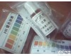
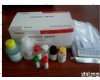
日本生研副流感病毒Ⅳ型PCR荧光试剂盒
广州健仑生物科技有限公司
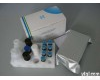
广州健仑长期供应各种PCR试剂盒,主要代理进口和国产品Pai的流行病毒PCR检测试剂盒。例如:甲乙型流感病毒核酸检测试剂盒、黄热病毒核酸检测试剂盒、诺如病毒核酸检测试剂盒、登革病毒核酸检测试剂盒、基孔肯雅病毒核酸检测试剂盒、结核杆菌核酸病毒检测试剂盒、孢疹病毒核算检测试剂盒、西尼罗河病毒PCR检测试剂盒、呼吸道合胞病毒核酸检测试剂盒、冠状病毒PCR检测试剂盒等等。虫媒体染病系列、呼吸道病原体系列、发热伴出疹系列、消化道及食源感染系列。
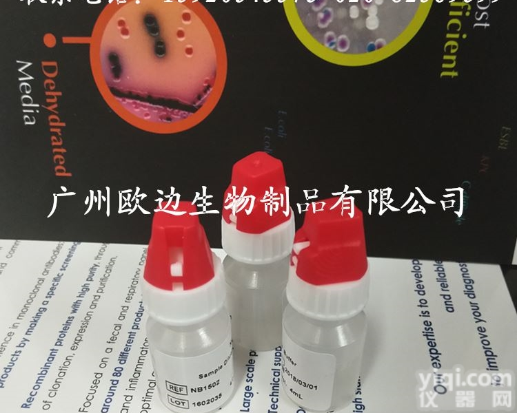
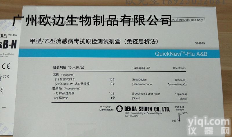
广州健仑长期供应各种流感检测试剂,包括进口和国产的品Pai,主要包括日本富士瑞必欧、日本生研、美国BD、美国NovaBios、美国binaxNOW、英国clearview、凯必利、广州创仑等主流品Pai。
主要检测:甲型流感病毒检测试剂、乙型流感病毒检测试剂、甲乙型流感病毒检测试剂、A+B流感病毒检测试剂盒、流感病毒抗原快速检测卡、流感病毒抗体快速检测试剂盒、流感快速检测试剂 c1c2。
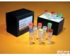
日本生研副流感病毒Ⅳ型PCR荧光试剂盒

我司还提供其它进口或国产试剂盒:登革热、疟疾、流感、A链球菌、合胞病毒、腮病毒、乙脑、寨卡、黄热病、基孔肯雅热、克锥虫病、违禁品滥用、肺炎球菌、军团菌、化妆品检测、食品安全检测等试剂盒以及日本生研细菌分型诊断血清、德国SiFin诊断血清、丹麦SSI诊断血清等产品。
欢迎咨询
欢迎咨询
想了解更多的产品及服务请扫描下方二维码:

【公司名称】 广州健仑生物科技有限公司
【市场部】 欧
【】
【腾讯 】
【公司地址】 广州清华科技园创新基地番禺石楼镇创启路63号二期2幢101-103室


巨大裂孔性视网膜脱离:巨大裂孔后瓣翻转、固定的巨大视网膜撕裂者,可予玻璃体切割、剥膜以解除玻璃体对视网膜的牵引。必要时作气体或硅油填充术和巩膜层间加压术。
视网膜僵硬或牵引性后极孔源性视网膜脱离:此类网脱病情复杂,常规手术操作困难,易损伤视神经及黄斑。可用玻璃体切割结合气体或硅油填充术ZL。
复发性视网膜脱离:由于多次常规网脱复位手术或玻璃体切割术的失败,导致了严重的玻璃体视网膜病变,视网膜固定皱褶多,网膜僵硬,活动性减弱。因此需作玻璃体切割及玻璃体填充联合巩膜环扎术。
视网膜脱离复位手术中有哪些并发症?
视网膜脱离复位手术中会出现一些并发症,常见的并发症有:①球壁穿孔。可发生在断腱、放水时。如穿孔发生在网膜脱离区,可作为放水孔处理;如发生在非网膜脱离区,应予缝合修补、局部冷凝及外加压。②放水并发症。除球壁穿孔外,如切口过大,液体流出过快,眼压骤降,可造成脉络膜渗出、出血,一旦发现即刻切开该处巩膜,放出脉络膜上腔的液体或血液,迅速结扎巩膜缝线及环扎条带。放水时过度压迫眼球可使视网膜、玻璃体球壁嵌顿,术后形成纤维血管膜,造成反复出血及牵引。③眼压升高:发生在脉络膜脱离时。宜予甘露醇静滴,必要时作前房穿刺。
视网膜脱离复位手术后有哪些并发症?
视网膜脱离复位手术中会出现并发症,手术后也会出现一些并发症,对于手术后的并发症也要引起重视。常见的术后并发症有以下几种。
(1)葡萄膜炎:视网膜手术因涉及葡萄膜,术后可发生葡萄膜炎。因此术后应局部或全身使用激素。
(2)眼内炎:较少见。可能通过放水口将病菌带入眼内。按常规处理,必要时作玻璃体切割。
(3)眼前段缺血:由于手术损伤睫状后长动脉或睫状前动脉后引起。轻度缺血较常见,不影响手术预后,重度缺血可造成严重后果,终致眼球萎缩。因此手术时宜少断直肌,3点与9点钟附近少电凝或冷凝,环扎不要太紧。ZL可全身或局部使用激素,必要时拆除环扎或加压物。
(4)视网膜下积液:可能是术中未放水,冷凝或电凝引起的渗出,裂孔封闭不良或遗漏及新的裂孔产生等。应作全面检查,渗出反应者全身或局部用激素,其他情况对症处理。
Large retinal detachment retinal detachment: a huge hole flap flap, a huge retinal tears were fixed, can be vitrectomy, stripping to lift the vitreous traction on the retina. If necessary, gas or silicone oil filling and scleral intercalation.
Retinal stiffness or traction Posterior rhegmatogenous retinal detachment: Such network disease complex, routine surgical difficulties, easy to damage the optic nerve and macula. Vitrectomy can be combined with gas or silicone oil filling treatment.
Recurrent retinal detachment: Due to the failure of many conventional mesh delaminating or vitrectomy, it leads to severe vitreoretinopathy, more fixed retinal folds, stiffer omentum, and weaker activity. Therefore, the need for vitrectomy and vitreous filling combined scleral cerclage. The company is located in:
What are the complications of retinal detachment surgery?
Retinal detachment surgery there will be some complications, the common complications are: ① perforation of the wall. Can occur in broken tendons, water when. Such as perforation occurred in the omentum detachment zone, can be treated as a drain hole; such as occurred in the non-retinal detachment area, should be sutured repair, local condensation and external pressure. ② drainage complications. In addition to perforation of the wall, such as incision is too large, the liquid flow too fast, sudden drop in intraocular pressure, can cause choroidal oozing, bleeding, once found immediay sclera, release the suprachoroidal fluid or blood, rapid ligation of the sclera Suture and ring tie strip. Excessive oppression when the eye drops can make the retina, vitreous wall incarcerated, the formation of fibrous vascular membrane, resulting in repeated bleeding and traction. ③ intraocular pressure: Occurred in the choroidal detachment. Should be mannitol intravenous infusion, if necessary, for the anterior chamber puncture. The company is located in:
What are the complications after retinal detachment surgery?
Retinal detachment surgery will appear complications in surgery, there will be some complications after surgery, complications for surgery should also pay attention. Common postoperative complications are the following.
(1) uveitis: retinal surgery involving the uvea, uveitis may occur after surgery. Therefore, postoperative hormones should be used locally or systemically.
(2) Endophthalmitis: less common. The bacteria may be brought into the eye through the drain. According to conventional treatment, if necessary, vitrectomy.
(3) anterior segment of ischemia: due to surgical damage caused by ciliary long artery or ciliary anterior artery caused. Mild ischemia is more common, does not affect the prognosis of surgery, severe ischemia can cause serious consequences, eventually causing the eye to shrink. Therefore, surgery should be less off rectus muscle, 3:00 and 9:00 less coagulation or condensation, cerclage not too tight. Treatment may be systemic or topical use of hormones, if necessary, removal of cerclage or pressure.
(4) subretinal fluid: the surgery may be no water, condensation or electrocoagulation caused by exudative, imperfect hole seal or missing new holes and so on. Should be fully inspected, exudation of systemic or topical response to hormones, symptomatic treatment in other cases.
本产品信息由(广州欧边生物制品有限公司)为您提供,内容包括(流感诊断试剂盒 日本生研副流感病毒Ⅳ型PCR荧光试剂盒)的品牌、型号、技术参数、详细介绍等;如果您想了解更多关于(流感诊断试剂盒 日本生研副流感病毒Ⅳ型PCR荧光试剂盒)的信息,请直接联系供应商,给供应商留言。若当前页面内容侵犯到您的权益,请及时告知我们,我们将马上修改或删除。
![[酶联免疫<em>试剂盒</em>] 猪ELISA<em>试剂盒</em> 猪瘟(CSF)ELISA<em>诊断试剂</em>盒](https://item.yiqi.com/pic/CovPic/1/20108411021175.jpg)